Odontogenic keratocyst involving zygoma: Cone-beam and Multislice spiral computed tomography aided the precise definition and appropriate treatment for the rare location
ID:16
Submission ID:35 View Protection:ATTENDEE
Updated Time:2021-10-30 07:09:50 Hits:1308
Poster Presentation

Start Time:2021-11-13 10:20 (Asia/Shanghai)
Duration:5min
Session:[Pos] Poster » [Pos] Poster Session
Video
No Permission
Presentation File
Tips: The file permissions under this presentation are only for participants. You have not logged in yet and cannot view it temporarily.
Abstract
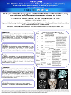
Background: Odontogenic keratocyst (OKC) has been reclassified back into the cystic category in 2017 WHO classification. However, it is uncommon OKC involved other bones except jaws. For superimposition of craniofacial structures in X-ray plain film and the rarity of OKC involving zygoma, diagnosis and treatment may be difficult in a certain degree.
Methods: The literature on OKCs involving zygoma was reviewed first. Then, retrospective research was carried out on the records of patients admitted to our hospital during a 36-year period. General clinic data, treatments, follow-up results and syndrome associated were analyzed.
Results: To the best of our knowledge, there has been only one reported case in the English literature since 1948. But 5 cases were found in our database. 4/5 patients were observed multiple lesions and suspected with Gorlin syndrome. All patients were treated by enucleation with curettage. Two of them experienced recurrence in the follow-up period ranging from 16 to 418 moths.
Conclusions: OKCs involving zygoma are really rare cases. Most patients with zygomatic involvements of OKCs displayed multiple lesions in both jaws and were suspected with Gorlin syndrome in this retrospective study. When patient was suspected with OKCs in maxilla, rational reserve of CT scanning was needed for excluding the extension into the zygoma. Cone-beam and multislice spiral computed tomography aided the precise definition and appropriate treatment for OKC involving the rare location zygoma.
Keywords
Odontogenic keratocyst; OKC; zygoma; Cone-beam computed tomography; CBCT; Multislice spiral computed tomography; MSCT
Speaker

Li Liu
Department of Oral Radiology, West China Hospital of Stomatology, State Key Laboratory of Oral Disease & National Clinical Research Center for Oral Disease, Sichuan University, Chengdu, PR China.Submission Author
All comments
Countdown
-
00
Days
-
00
Hours
-
00
Minutes
-
00
Seconds
Important Dates
| Abstract submission date: |
2021-10-25 |
| Full paper submission date: |
2021-10-25 |
| Notification of acceptance date: |
2021-11-01 |
| Conference date: | 2021-11-12~2021-11-14 |
Contact Us
| Jinying Yang | +86 13675518597 |
| Debo Zhi | +86 15056085235 |
| Song Gao | +86 13121880288 |
| Le Cao | +86 15910809908 |
Comment submit